
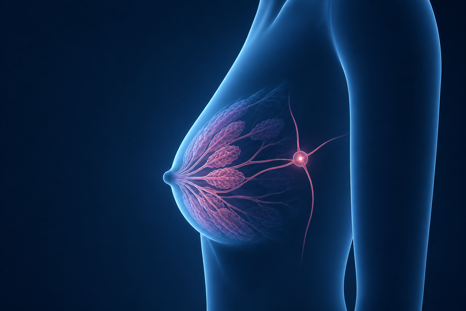
Selected examples from the broader physician-developed education library available through an UnderstandMyScan breast center partnership.
Breast density refers to the proportion of fibroglandular tissue (glands and connective tissue) compared to fatty tissue on a mammogram. Dense breasts have more fibroglandular tissue and are extremely common - roughly 40-50% of women have heterogeneously or extremely dense breasts.
Dense tissue has two important implications. First, it can partially obscure small masses on a mammogram because both dense tissue and tumors appear white on the image. Second, dense breasts carry a modestly elevated independent risk for breast cancer. Many states now require that patients be notified if their breasts are dense, and supplemental screening with ultrasound or MRI may be recommended.
Deep Dive: Dense Breast Tissue ExplainedA callback (also called a recall) after a screening mammogram is very common and is usually not cause for alarm. Approximately 10-12% of women are called back after their first mammogram, and roughly 5-10% after subsequent screenings. The large majority of callbacks result in a normal outcome.
A callback means the radiologist saw something that needs a closer look - often an asymmetry, a possible mass, or calcifications that are easier to see with additional views or ultrasound. A BI-RADS 0 is assigned, which means the study is incomplete pending further imaging. This is a diagnostic process, not a diagnosis of cancer.
Deep Dive: Understanding BI-RADS ScoresCalcifications are tiny calcium deposits in breast tissue that show up as bright white specks or clusters on a mammogram. They are extremely common and the majority are completely benign - caused by aging, past injury, or normal cellular activity.
What matters to a radiologist is the size, shape, and distribution of the calcifications. Large, round, smooth calcifications are almost always benign. Small, irregular, or tightly clustered (especially in a line) calcifications may require biopsy because they can occasionally be associated with early breast cancer or a precancerous condition called DCIS. Your radiologist will assign a BI-RADS score based on this assessment.
Deep Dive: Breast Calcifications ExplainedBI-RADS 4 means a finding looks suspicious enough that a biopsy is usually recommended. It does not mean you definitely have breast cancer. It means imaging cannot confidently call the area benign, so tissue sampling is recommended to get a clear answer. BI-RADS 4 covers a wide range of concern, from low suspicion to higher suspicion.
Deep Dive: BI-RADS 4A breast biopsy is a procedure in which a small sample of tissue is removed from the suspicious area and examined by a pathologist under a microscope. It is the only way to know definitively whether a finding is benign or malignant. Being recommended for biopsy does not mean you have cancer - the majority of biopsied lesions turn out to be benign.
Most breast biopsies today are performed as minimally invasive image-guided procedures using a hollow needle - either ultrasound-guided or stereotactic (X-ray-guided). The procedure is done under local anesthesia, takes about 30-60 minutes, and does not require surgery. A small clip is typically placed at the biopsy site as a marker. Results are usually available within 2-3 business days.
Deep Dive: What to Expect From a Breast BiopsyThe full partner platform includes a broader patient education library, additional FAQs, staff communication tools, and workflow-connected resources customized for each breast center.
Explore a Partnership